A Playful Aerobic Microbe Sterility “Test”

HouseCat

Explorer
Joined
May 31, 2026
Messages
434
Reaction score
0
Location
New York
I picked up some agar plates so I could check how sterile my peptide solutions are and carry out an “experiment.” I put experiment in quotation marks since it can’t really be repeated exactly, though it’s a bit more than a simple project!

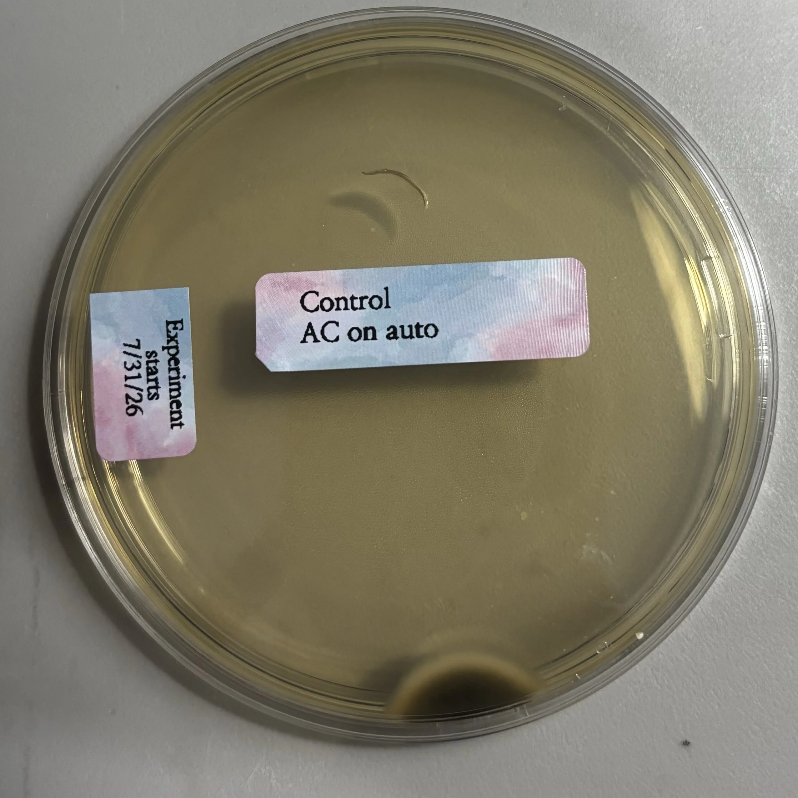
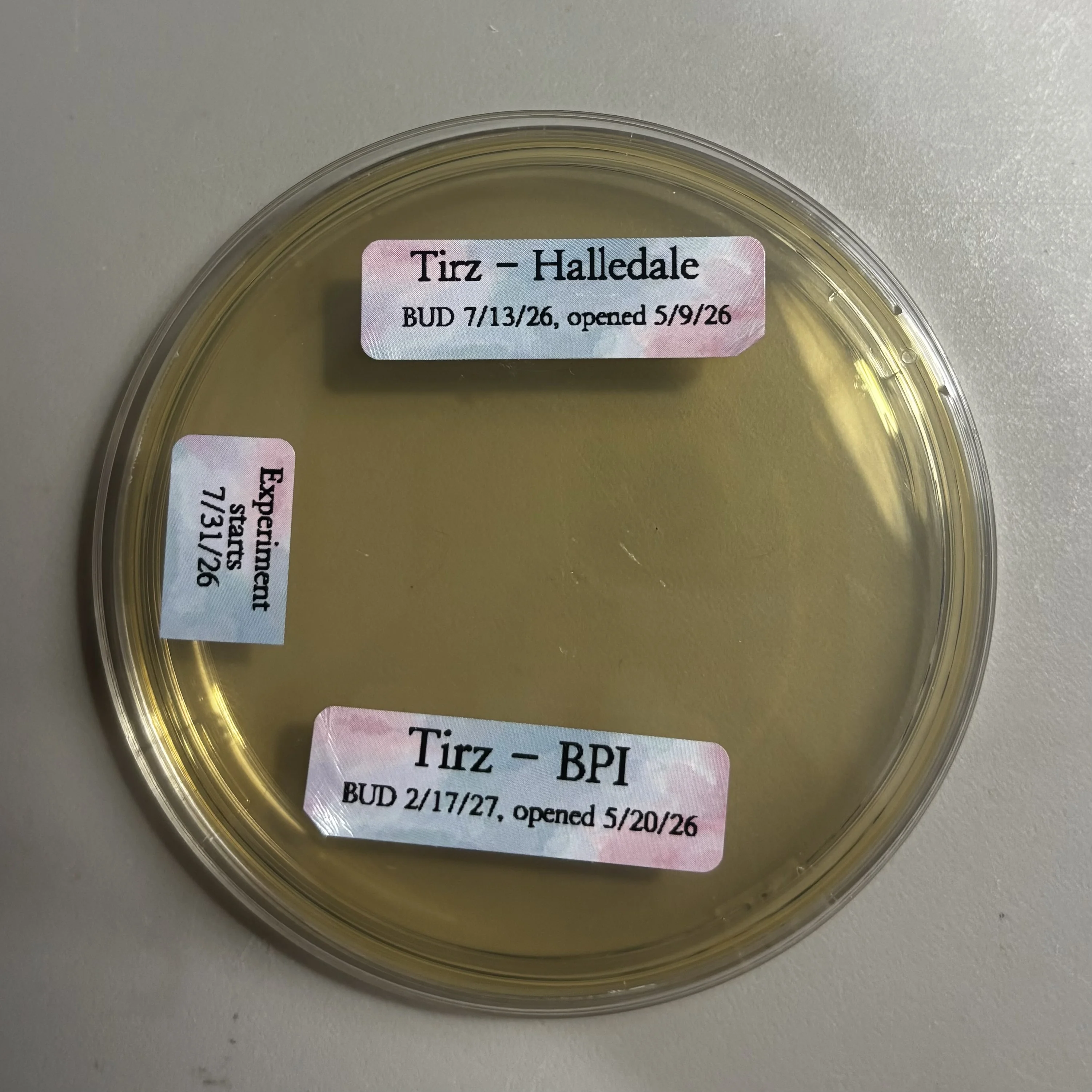
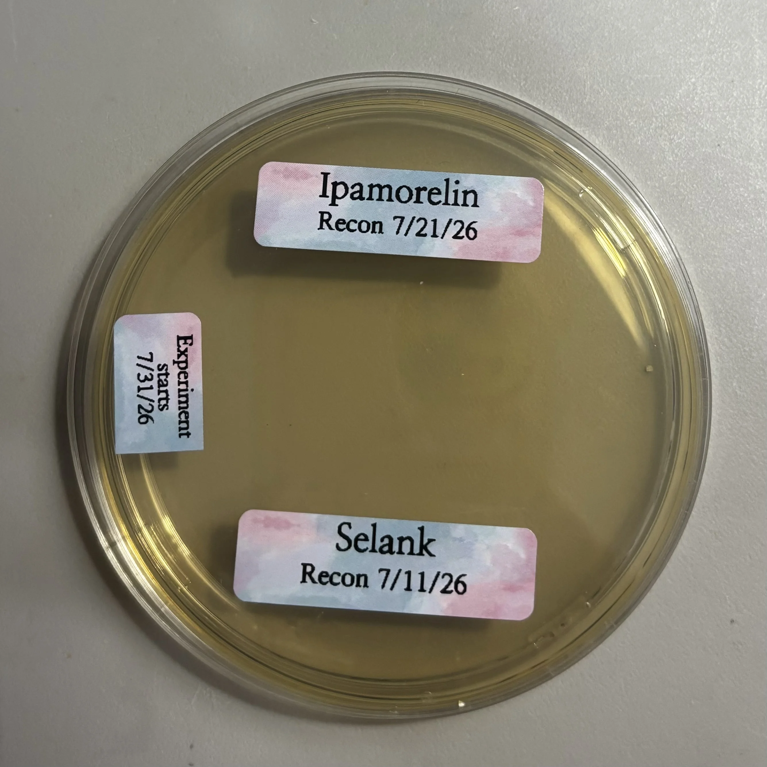
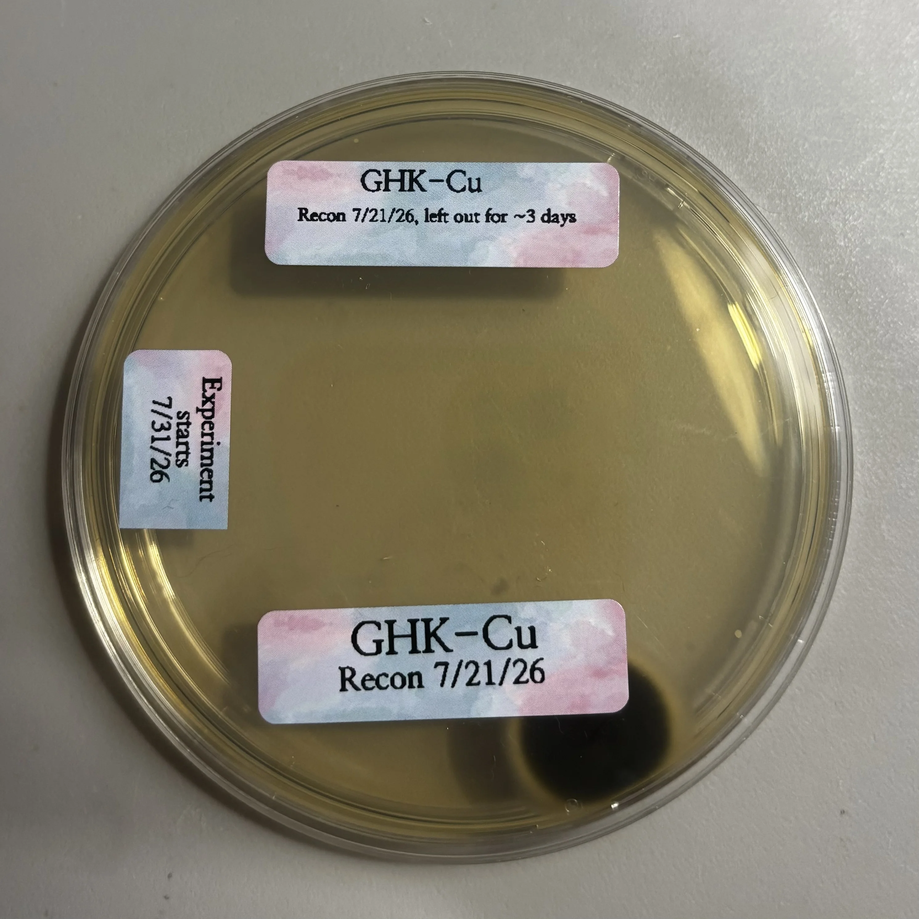
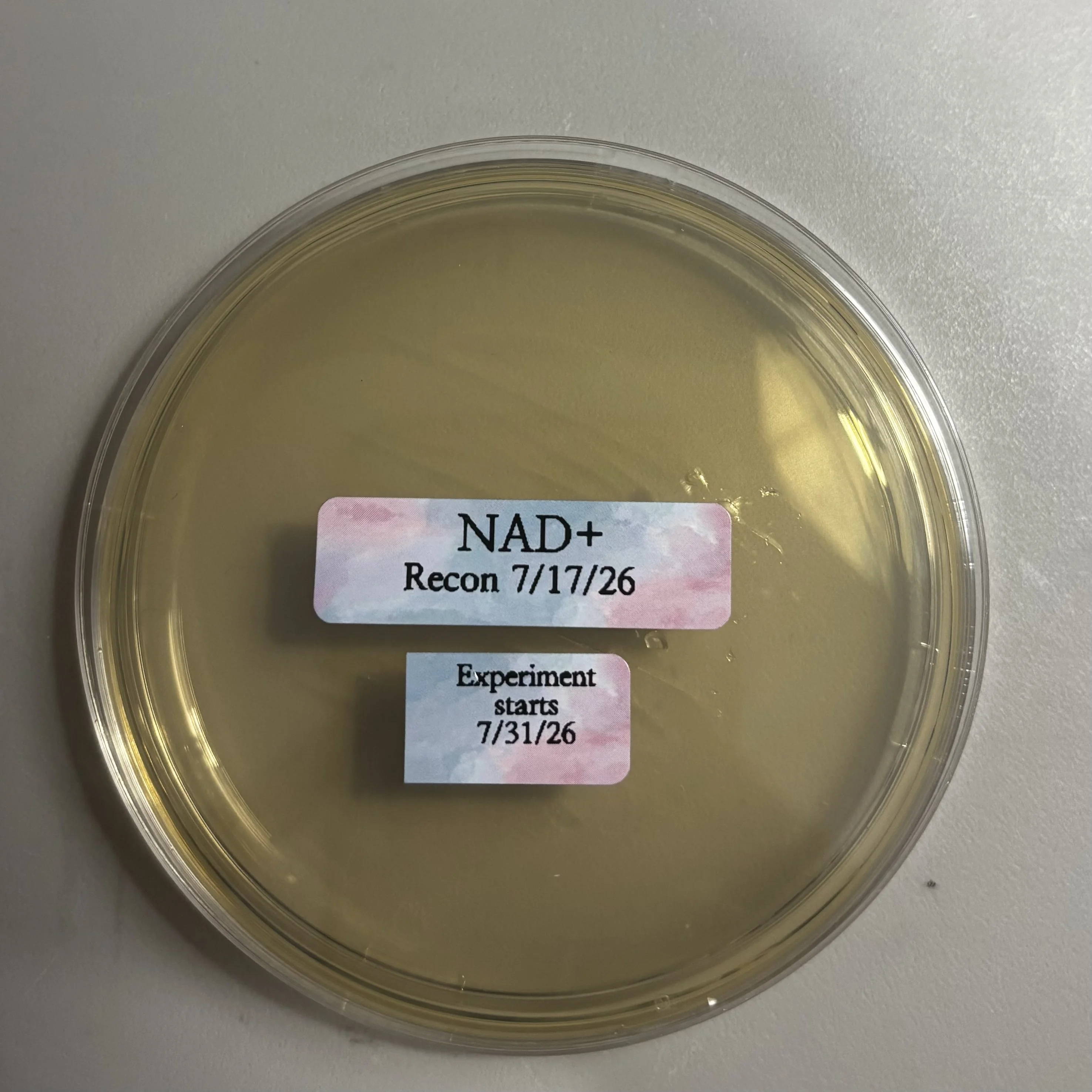
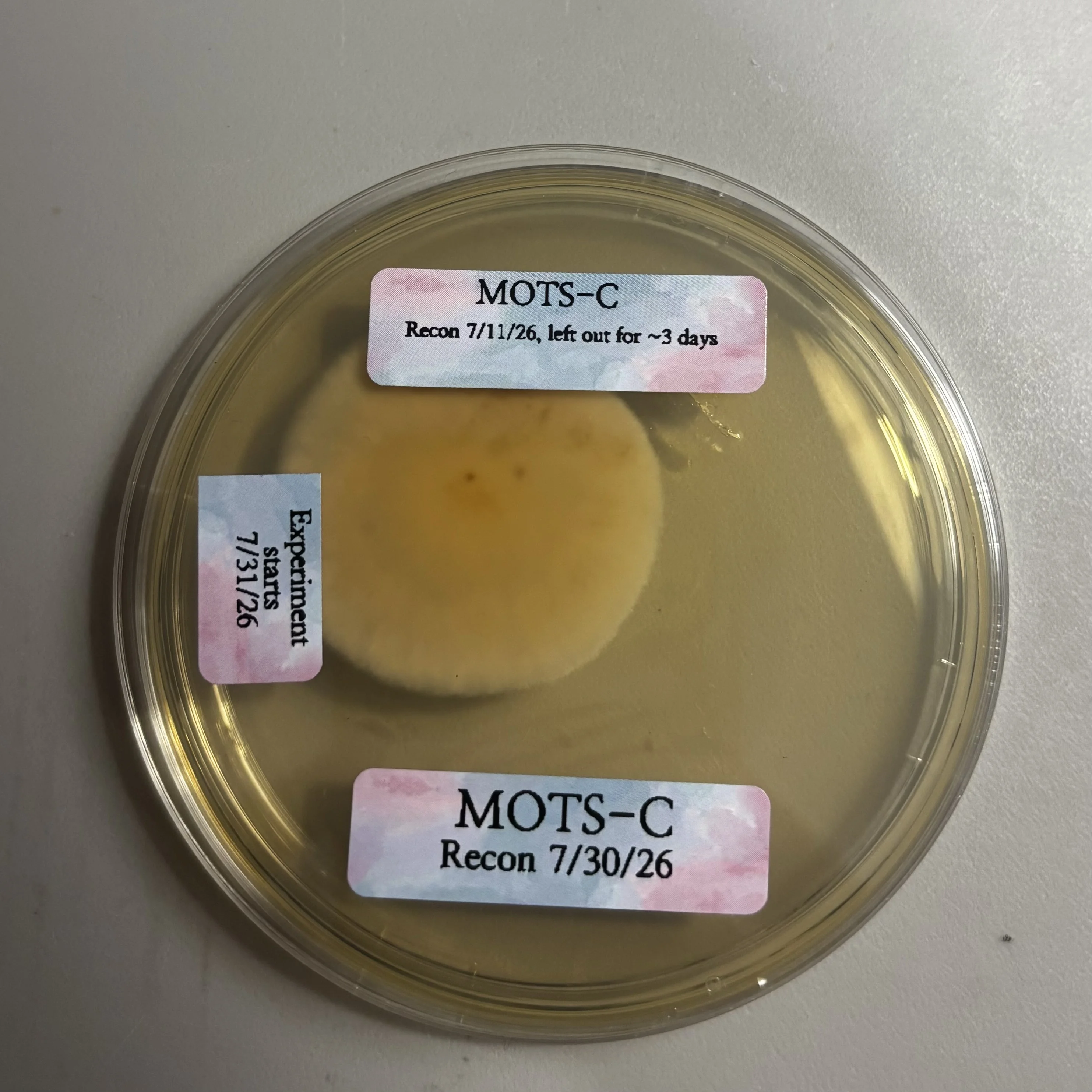
How I did it: using either sterile pen needles or sterile syringes, I deposited roughly 1 unit of solution onto the agar plates; every solution had its own sterile needle/syringe. I tried to land about 2.5cm in from the outer edge, then spread the solution across an area of roughly 3x2cm with sterile, individually wrapped cotton swabs. My AC ran throughout, set to auto (so sometimes it blew, sometimes it didn’t). The plates stayed covered and incubated in a room that holds around 70F. Every plate was left open to the air for 2.5 to 5.5 minutes, and the control was exposed for the same length of time as the longest-exposed experimental plate. (A few solutions were being used at the time, so I’d put solution on the plate with a sterile needle, then inject myself, which explains the timing differences. Selank and Ipamorelin were handled that way and shared a plate, so it took a bit longer.) MOTS-C and GHK-Cu each had fresh/refrigerated samples plus samples from the leftovers of cartridges that had sat at room temp for 3 days. Refrigerated cartridge samples (not tirz, which was in vials) had been used for double needle (standard pen needles) injections before testing. (The BPI had its cap taken off on the labeled date but wasn’t pierced until many weeks afterward, though I don’t remember when. Once I got into gray, I decided I could afford to play around with my compounded to figure out which formula I prefer; it’s BPI.)

Problems: The desk I first used was not level, so Selank and Ipamorelin and the refrigerated GHK-Cu samples ran along the edge into the other solution’s area; after that I moved to a level desk. The pen put out a lot of force, so the solution splattered more than with the syringe. I ended up touching the control sample with my bare fingers in the middle of the plate because I thought the plate was still covered—you can see where my nail pierced the agar. The cotton swabs had a pointy end, which I used, but on the NAD+ plate I pressed too hard and scratched the agar.

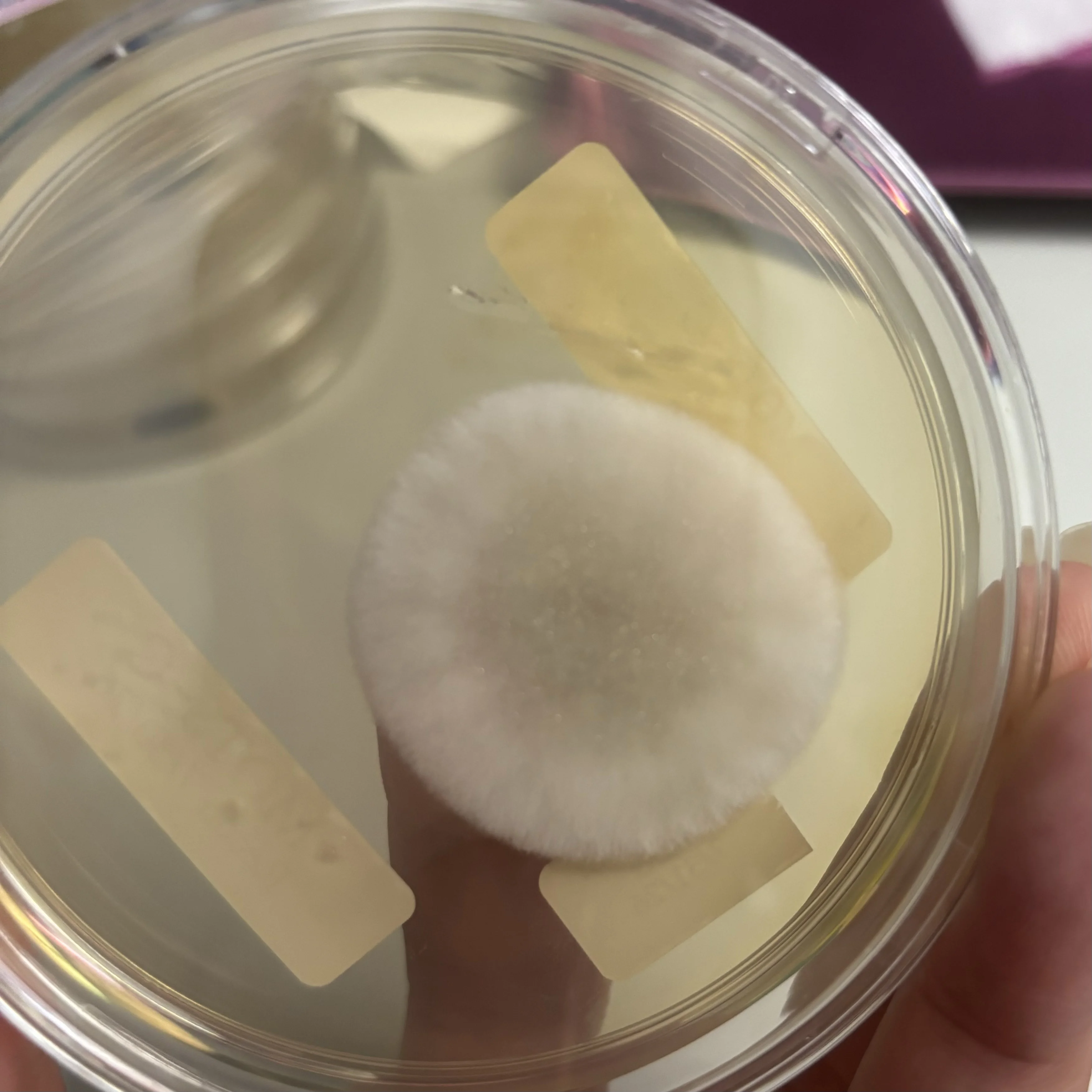
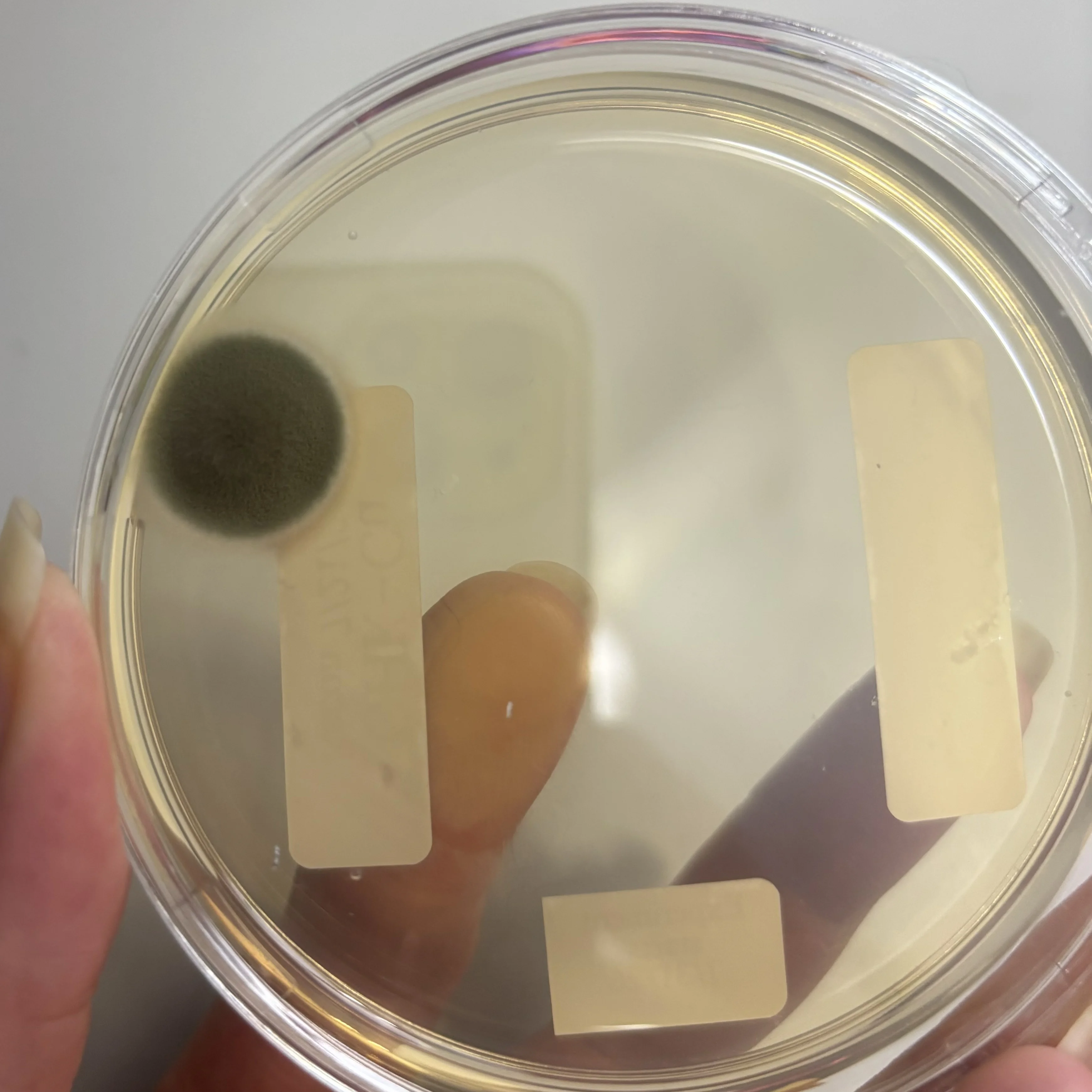
Results: Mostly good! Some inconclusive, some fairly conclusive. No growth where I touched the control plate: my hand hygiene is on point-woo hoo! The Control sample and the GHK-Cu both show what looks like the same growth at the edge of the plate. That makes me think it came from directional airflow from my AC and was something in the air rather than unsterile GHK-Cu, especially since I don’t see growth where I placed the actual samples. NAD+ is inconclusive…I’m not sure whether what I’m seeing is scratches or growth, since I don’t have a microscope and to my eye it looks the same as when I first spread the sample. MOTS-C that had been left out for 3 days…whoo! There is something really funky there, and I’m pretty sure it’s not from airflow given the placement. (Yes, I had injected this before leaving the sample out…no ill effects except possibly a multi-day itchy ISR, but I can’t exactly recall what pin it was from and when.)

Next month I will test different samples for anaerobic bacteria, and in the fall I will test for aerobic bacteria on blood agar plates.
d20f42246ad1360f991cec083ae2b44500ffb3e63f132ca14d66660e59103c49.webp


dd727ab484449eb6958fb5bfa6c1c89239377f8cbff63f05cc4d29b9ebd3e36c.webp


f8cba0b2aba096aea9d7a90bfc4bf4dd85960043d4613825a111671e75067ef0.webp


104ac8507604a6372810ee1172ac07e6efc5de435d2b0aca77f7cbd0217446b8.webp


a92921538c314dce61c46e74908f29fc9b32e8d7f6ffed8035a089127b5aa0e5.webp


aac3c233541a045127499d09aa47a78cea768d648f70b4cfedec27cffc130bf5.webp


MOTS-C other side

255d927665b1051127a52615b203da0129d5250a656dfe690a46b7f34592a3c7.webp


GHK-Cu, other side

24f560ff4e55b0640691ce2cf4a8a7ea117b086fc2ae40b53cbe03794b1a6c23.webp
 
What a blast!

There’s a lot I want to ask.. but I’ll return once this thread has been live for a while.

Appreciate you putting this together!!
 
Wow! That seems like a good time.

On Peppy's, I saw a post where somebody tried something comparable, though they used vials and checked for cloudiness as time passed. Really fascinating.

I appreciate you posting this.
 
Much appreciated. I have a lot of respect for folks who run this kind of test and then share what they find — be it checking for bacteria on an agar plate or self-administering unusual compounds or amounts.
 
Grogu said:

Wow! That seems like a good time.

On Peppy's, I saw a post where somebody tried something comparable, though they used vials and checked for cloudiness as time passed. Really fascinating.

I appreciate you posting this.
I caught that too! And that one’s headed for the plate…😂 hold on—the vial—next month! I’ll be working with liquid growth medium sourced from Biologix.
 
Good to know — I’d been curious whether anybody had tried something along these lines! If my understanding were stronger, I’d pitch in and lend a hand.
 
That appears to be a round, white, filamentous fungal colony. A single spore is enough. Maybe it was already inside the vial, or maybe it was airborne and got sucked into the syringe as you drew the plunger down. It's also possible it just happened to land on the plate. If the vial itself were the source, I'd anticipate seeing contamination in that vial as well.
 
Just A Cat said:

That appears to be a round, white, filamentous fungal colony. A single spore is enough. Maybe it was already inside the vial, or maybe it was airborne and got sucked into the syringe as you drew the plunger down. It's also possible it just happened to land on the plate. If the vial itself were the source, I'd anticipate seeing contamination in that vial as well.
Yep, every bit of that checks out! Nobody can say with certainty either way. My plan was to leave the AC running the whole time, just to find out whether that would hurt sterility for every plate. If I were being rigorous, though, I'd set up a comparison between AC air and a still air box across every sample. Realistically that won't happen—this “experiment” is a low-effort thing for me.

After sitting out for 3 day, the MOTS-C vial had gone cloudy, and I only caught it when the cartridge needed swapping. I'd gotten careless about checking carts before pinning, and that won't be happening again. The replacement MOTS-C went cloudy 1 day post-recon too, though. Saline BAC was used for both, and I'm done using it with MOTS-C given that both vials clouded up. An earlier MOTS-C (same store and supplier—CytoBlaze, different mg, every vial tested well & filtered) was mixed with plain BAC and stayed clear, even though it foamed a bit at first.

BTW, every BAC/NaCl BAC was Hospira.

EDIT: On “experiment” day the newer MOTS-C was still clear, so the cloudiness must have set in soon afterward (sometime 8/1 or later, before 8/4 when I spotted it). Whether that cloudiness comes from a reaction to Saline or from contamination, I can't say. A chemical reaction ought to show up faster, I'd think, but chem isn't my strong suit. ( @eidos if you want to weigh in, much appreciated! ) I also don't have enough MOTS-C to spare for testing another MOTS-C + NaCl BAC from a different vendor LOL! For this pep I'm a single vial gal until my kit order lands. Which bums me out, because the NaCl MOTS (from the older vial) was defs less spicy than MOTS+BAC.
 
Girllll, you never stop surprising me. That is some seriously impressive work!
 
From what I’ve seen, edge contamination tends to come from inadequate wrapping or a parafilm breakdown. Using the middle portion of the plate, rather than the outer rim, is always the better approach.

What kind of wrap are you using? I gave one brand a shot, and it was terrible (it wouldn’t stretch right and had little rips). Once I switched back to genuine parafilm, the problems stopped.
 
CandyCap said:

From what I’ve seen, edge contamination tends to come from inadequate wrapping or a parafilm breakdown. Using the middle portion of the plate, rather than the outer rim, is always the better approach.

What kind of wrap are you using? I gave one brand a shot, and it was terrible (it wouldn’t stretch right and had little rips). Once I switched back to genuine parafilm, the problems stopped.
Nothing was used to wrap them, so contamination at the edges caused by not wrapping is definitely possible! I appreciate you pointing that out—I’ll make sure to wrap the blood agar plates when I use them. That step had completely slipped my mind…for me microbio was around 10 years ago, so my lab procedures and precautions aren’t as sharp as they are for those of you doing mushroom and plant propagation!

I put the samples a few cm in from the edges, but the uneven desk and the whole “liquids go downhill” thing caused problems there 😆 I also put two samples on several plates because I wanted to test as much as possible without buying additional plates—again not best practice, but definitely fun (especially seeing them come out clear, well that’s more of a relief… the growths are less of a relief but pretty fun!)
 
None of this involved proper laboratory-grade clinical testing. Every one of your workarounds looks doable. And enjoyable!
 
Just A Cat said:

None of this involved proper laboratory-grade clinical testing. Every one of your workarounds looks doable. And enjoyable!
100%!! Definitely not clinical in any way, far too hard to reproduce, plenty of looseness, and mostly built around keeping my in-use peps as cold as I can while playing around with light science. And yep, 100% fun too! 🤩
 
HouseCat said:

CandyCap said:

From what I’ve seen, edge contamination tends to come from inadequate wrapping or a parafilm breakdown. Using the middle portion of the plate, rather than the outer rim, is always the better approach.

What kind of wrap are you using? I gave one brand a shot, and it was terrible (it wouldn’t stretch right and had little rips). Once I switched back to genuine parafilm, the problems stopped.
Nothing was used to wrap them, so contamination at the edges caused by not wrapping is definitely possible! I appreciate you pointing that out—I’ll make sure to wrap the blood agar plates when I use them. That step had completely slipped my mind…for me microbio was around 10 years ago, so my lab procedures and precautions aren’t as sharp as they are for those of you doing mushroom and plant propagation!

I put the samples a few cm in from the edges, but the uneven desk and the whole “liquids go downhill” thing caused problems there 😆 I also put two samples on several plates because I wanted to test as much as possible without buying additional plates—again not best practice, but definitely fun (especially seeing them come out clear, well that’s more of a relief… the growths are less of a relief but pretty fun!)
When the syringe is being stubborn, rather than pushing the plunger, try pressing on the barrel itself. That way you get somewhat finer control over the flow 🙂 A couple of drops ought to be plenty, too.

My habit is to work only within the middle 2/3rds of the plate.
 
CandyCap said:

HouseCat said:

CandyCap said:

From what I’ve seen, edge contamination tends to come from inadequate wrapping or a parafilm breakdown. Using the middle portion of the plate, rather than the outer rim, is always the better approach.

What kind of wrap are you using? I gave one brand a shot, and it was terrible (it wouldn’t stretch right and had little rips). Once I switched back to genuine parafilm, the problems stopped.
Nothing was used to wrap them, so contamination at the edges caused by not wrapping is definitely possible! I appreciate you pointing that out—I’ll make sure to wrap the blood agar plates when I use them. That step had completely slipped my mind…for me microbio was around 10 years ago, so my lab procedures and precautions aren’t as sharp as they are for those of you doing mushroom and plant propagation!

I put the samples a few cm in from the edges, but the uneven desk and the whole “liquids go downhill” thing caused problems there 😆 I also put two samples on several plates because I wanted to test as much as possible without buying additional plates—again not best practice, but definitely fun (especially seeing them come out clear, well that’s more of a relief… the growths are less of a relief but pretty fun!)
When the syringe is being stubborn, rather than pushing the plunger, try pressing on the barrel itself. That way you get somewhat finer control over the flow 🙂 A couple of drops ought to be plenty, too.

My habit is to work only within the middle 2/3rds of the plate.
Those are useful pointers… what’s the technique for squeezing the syringe? The ones I have are fairly stiff plastic, though I admit I’ve never actually attempted to squeeze them. I only went through roughly 1 unit, so how would I manage to get a grip on it beneath the plunger—or does squeezing above it do the job too? Really intriguing!

Honestly, I’ll probably run two samples on each blood agar plate because it’s quite expensive, but who knows? Maybe I’ll hit it big on a scratcher!…If I actually played 😂 or perhaps a Bank error in my favor? 🤣

EDIT: By the way, I’m using 0.3ml syringes, brand unknown. I also have 1mls, but the smaller ones make units easier to see.
 
Grogu said:

Wow! That seems like a good time.

On Peppy's, I saw a post where somebody tried something comparable, though they used vials and checked for cloudiness as time passed. Really fascinating.

I appreciate you posting this.
The results are out from jonny /community-link/thread/20630/
 
HouseCat said:

CandyCap said:

HouseCat said:

CandyCap said:

From what I’ve seen, edge contamination tends to come from inadequate wrapping or a parafilm breakdown. Using the middle portion of the plate, rather than the outer rim, is always the better approach.

What kind of wrap are you using? I gave one brand a shot, and it was terrible (it wouldn’t stretch right and had little rips). Once I switched back to genuine parafilm, the problems stopped.
Nothing was used to wrap them, so contamination at the edges caused by not wrapping is definitely possible! I appreciate you pointing that out—I’ll make sure to wrap the blood agar plates when I use them. That step had completely slipped my mind…for me microbio was around 10 years ago, so my lab procedures and precautions aren’t as sharp as they are for those of you doing mushroom and plant propagation!

I put the samples a few cm in from the edges, but the uneven desk and the whole “liquids go downhill” thing caused problems there 😆 I also put two samples on several plates because I wanted to test as much as possible without buying additional plates—again not best practice, but definitely fun (especially seeing them come out clear, well that’s more of a relief… the growths are less of a relief but pretty fun!)
When the syringe is being stubborn, rather than pushing the plunger, try pressing on the barrel itself. That way you get somewhat finer control over the flow 🙂 A couple of drops ought to be plenty, too.

My habit is to work only within the middle 2/3rds of the plate.
Those are useful pointers… what’s the technique for squeezing the syringe? The ones I have are fairly stiff plastic, though I admit I’ve never actually attempted to squeeze them. I only went through roughly 1 unit, so how would I manage to get a grip on it beneath the plunger—or does squeezing above it do the job too? Really intriguing!

Honestly, I’ll probably run two samples on each blood agar plate because it’s quite expensive, but who knows? Maybe I’ll hit it big on a scratcher!…If I actually played 😂 or perhaps a Bank error in my favor? 🤣

EDIT: By the way, I’m using 0.3ml syringes, brand unknown. I also have 1mls, but the smaller ones make units easier to see.
Overfill the syringe—more than the amount you actually need. My choice is 3ml luer locks.

For my own plates, I usually pour MEA, PDA, or just water plates (these last ones work well to clean up contaminants), and it’s fairly cheap.

Blood plates are outside what I can advise on....right now, the very last thing this world needs is me tinkering with blood and mycelium... that could turn into The Last of Us
 
HouseCat said:

I picked up some agar plates so I could check how sterile my peptide solutions are and carry out an “experiment.” I put experiment in quotation marks since it can’t really be repeated exactly, though it’s a bit more than a simple project!

How I did it: using either sterile pen needles or sterile syringes, I deposited roughly 1 unit of solution onto the agar plates; every solution had its own sterile needle/syringe. I tried to land about 2.5cm in from the outer edge, then spread the solution across an area of roughly 3x2cm with sterile, individually wrapped cotton swabs. My AC ran throughout, set to auto (so sometimes it blew, sometimes it didn’t). The plates stayed covered and incubated in a room that holds around 70F. Every plate was left open to the air for 2.5 to 5.5 minutes, and the control was exposed for the same length of time as the longest-exposed experimental plate. (A few solutions were being used at the time, so I’d put solution on the plate with a sterile needle, then inject myself, which explains the timing differences. Selank and Ipamorelin were handled that way and shared a plate, so it took a bit longer.) MOTS-C and GHK-Cu each had fresh/refrigerated samples plus samples from the leftovers of cartridges that had sat at room temp for 3 days. Refrigerated cartridge samples (not tirz, which was in vials) had been used for double needle (standard pen needles) injections before testing. (The BPI had its cap taken off on the labeled date but wasn’t pierced until many weeks afterward, though I don’t remember when. Once I got into gray, I decided I could afford to play around with my compounded to figure out which formula I prefer; it’s BPI.)

Problems: The desk I first used was not level, so Selank and Ipamorelin and the refrigerated GHK-Cu samples ran along the edge into the other solution’s area; after that I moved to a level desk. The pen put out a lot of force, so the solution splattered more than with the syringe. I ended up touching the control sample with my bare fingers in the middle of the plate because I thought the plate was still covered—you can see where my nail pierced the agar. The cotton swabs had a pointy end, which I used, but on the NAD+ plate I pressed too hard and scratched the agar.

Results: Mostly good! Some inconclusive, some fairly conclusive. No growth where I touched the control plate: my hand hygiene is on point-woo hoo! The Control sample and the GHK-Cu both show what looks like the same growth at the edge of the plate. That makes me think it came from directional airflow from my AC and was something in the air rather than unsterile GHK-Cu, especially since I don’t see growth where I placed the actual samples. NAD+ is inconclusive…I’m not sure whether what I’m seeing is scratches or growth, since I don’t have a microscope and to my eye it looks the same as when I first spread the sample. MOTS-C that had been left out for 3 days…whoo! There is something really funky there, and I’m pretty sure it’s not from airflow given the placement. (Yes, I had injected this before leaving the sample out…no ill effects except possibly a multi-day itchy ISR, but I can’t exactly recall what pin it was from and when.)

Next month I will test different samples for anaerobic bacteria, and in the fall I will test for aerobic bacteria on blood agar plates.


View attachment 9927

View attachment 9928

View attachment 9929

View attachment 9930

View attachment 9931

View attachment 9932

MOTS-C other side

View attachment 9933

GHK-Cu, other side

View attachment 9934
These are pretty neat experiments. I might have missed it, but did you say whether you ran the peptides through a filter during reconstitution? I wasn't sure if that step came before the testing.
 
My knowledge of microbes isn't deep enough to give a definite answer, though I've watched quite a few of these tests run on our machines. What's visible here resembles mold rather than bacteria. Naturally, confirming that would require a microscope, yet the filaments and the fuzzy texture are plainly apparent.
 
HouseCat said:

I picked up some agar plates so I could check how sterile my peptide solutions are and carry out an “experiment.” I put experiment in quotation marks since it can’t really be repeated exactly, though it’s a bit more than a simple project!

How I did it: using either sterile pen needles or sterile syringes, I deposited roughly 1 unit of solution onto the agar plates; every solution had its own sterile needle/syringe. I tried to land about 2.5cm in from the outer edge, then spread the solution across an area of roughly 3x2cm with sterile, individually wrapped cotton swabs. My AC ran throughout, set to auto (so sometimes it blew, sometimes it didn’t). The plates stayed covered and incubated in a room that holds around 70F. Every plate was left open to the air for 2.5 to 5.5 minutes, and the control was exposed for the same length of time as the longest-exposed experimental plate. (A few solutions were being used at the time, so I’d put solution on the plate with a sterile needle, then inject myself, which explains the timing differences. Selank and Ipamorelin were handled that way and shared a plate, so it took a bit longer.) MOTS-C and GHK-Cu each had fresh/refrigerated samples plus samples from the leftovers of cartridges that had sat at room temp for 3 days. Refrigerated cartridge samples (not tirz, which was in vials) had been used for double needle (standard pen needles) injections before testing. (The BPI had its cap taken off on the labeled date but wasn’t pierced until many weeks afterward, though I don’t remember when. Once I got into gray, I decided I could afford to play around with my compounded to figure out which formula I prefer; it’s BPI.)

Problems: The desk I first used was not level, so Selank and Ipamorelin and the refrigerated GHK-Cu samples ran along the edge into the other solution’s area; after that I moved to a level desk. The pen put out a lot of force, so the solution splattered more than with the syringe. I ended up touching the control sample with my bare fingers in the middle of the plate because I thought the plate was still covered—you can see where my nail pierced the agar. The cotton swabs had a pointy end, which I used, but on the NAD+ plate I pressed too hard and scratched the agar.

Results: Mostly good! Some inconclusive, some fairly conclusive. No growth where I touched the control plate: my hand hygiene is on point-woo hoo! The Control sample and the GHK-Cu both show what looks like the same growth at the edge of the plate. That makes me think it came from directional airflow from my AC and was something in the air rather than unsterile GHK-Cu, especially since I don’t see growth where I placed the actual samples. NAD+ is inconclusive…I’m not sure whether what I’m seeing is scratches or growth, since I don’t have a microscope and to my eye it looks the same as when I first spread the sample. MOTS-C that had been left out for 3 days…whoo! There is something really funky there, and I’m pretty sure it’s not from airflow given the placement. (Yes, I had injected this before leaving the sample out…no ill effects except possibly a multi-day itchy ISR, but I can’t exactly recall what pin it was from and when.)

Next month I will test different samples for anaerobic bacteria, and in the fall I will test for aerobic bacteria on blood agar plates.


View attachment 9927

View attachment 9928

View attachment 9929

View attachment 9930

View attachment 9931

View attachment 9932

MOTS-C other side

View attachment 9933

GHK-Cu, other side

View attachment 9934
Watch your back, Jano — a cat is moving in on your turf.
 
Back
Top